General
We book you in our next available appointment opening.
X-ray
Ultrasound
CT (Computed Tomography)
Our CT is a separate department that accepts CT patients under contract with the Regina Qu’Appelle Health Region. All referrals go through the region, the same as other CT requests, and the region then distribute the referrals to the facilities that do the exams.
Yes, ultimately it is your right to accept or decline to have the contrast. Please keep in mind that the Radiologist may not be able to provide your physician with as much information without contrast as they might have been able to, had you had the contrast. Your exam may be cancelled as the Radiologist will not be able to answer the question your physician is trying to get an answer about. In some instances an alternative exam may be your best choice; therefore your exam may need to be rescheduled as we may need to prepare you for the test differently.
No that was most likely a MRI. During your CT scan you will hear the humming of the machine but it is not as loud as the MRI machines.
Yes, your doctor must send a referral to the Regina General Hospital, in order for you to receive a computed tomography (CT) imaging examination. However, the Regina Hospital may book outpatient CT exams at Prairie Skies Medical Imaging Parliament Ave location.
Your exam will be read by a Radiologist: a Physician specialist in diagnostic imaging. The radiologist will analyze the images and send an official report to the physician that referred you for the exam, who will then discuss the results with you.
In many cases, you will not meet the radiologist who is working on your behalf. You can trust, however, that they have the knowledge and experience to help you and your doctor make informed decisions about your health care.
No, but if you do not speak fluent English please bring someone who is can help to translate any instructions.
Typically, family members or friends, especially children are not permitted to stay with you in the CT room. CT scanners use ionizing radiation and only the patient requiring the scan is permitted in the room. With pediatric patients, a parent may be allowed in the room but will be required to wear a lead apron to minimize radiation exposure. Do not bring pets, as there is no supervision for them during your test.
Pregnant woman should not have a CT exam or any x-ray examination, especially if the woman is in her first trimester (first of three-3 month periods of pregnancy). Depending on the condition, there may be other exams available, such as ultrasound, to help diagnose a medical condition. Pregnant women should always inform their imaging technologist or radiologist that they are pregnant, or may be pregnant.
It is natural to have questions or concerns about your CT scan. Please don't hesitate to discuss them with your doctor or specialist.
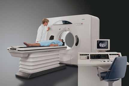
 The CT scanner is typically a large, box-like machine with a hole, or short tunnel, in the center. You will lie on a narrow examination table that slides into and out of this tunnel. Rotating around you, the x-ray tube and electronic x-ray detectors are located opposite each other in a ring, called a gantry. The computer workstation that processes the imaging information is located in a separate control room, where the technologist operates the scanner and monitors your examination in direct visual contact and usually with the ability to hear and talk to you with the use of a speaker and microphone.
Modern CT scanners are so fast that they can scan through large sections of the body in just a few seconds, and even faster in small children. Such speed is beneficial for all patients but especially children, the elderly and critically ill, all of whom may have difficulty in remaining still, even for the brief time necessary to obtain images.
When you arrive for your appointment, a technologist will take you to the CT scanning room, where you will see a table and a large doughnut-shaped device, called a gantry. Our technologist will ask you to lie on the padded table and make sure you're comfortable. The table can be raised or lowered. Straps and pillows may be used to help you maintain the correct position and to help you remain still during the exam. During a CT scan of the head, the table may be fitted with a special cradle that holds your head still. Next, the table will move quickly through the scanner to determine the correct starting position for the scans. Then, the table will move slowly through the machine as the actual CT scanning is performed. Depending on the type of CT scan, the machine may make several passes.
You may be asked to hold your breath during the scanning. Any motion, whether breathing or body movements, can lead to artifacts on the images. This loss of image quality can resemble the blurring seen on a photograph taken of a moving object.
During the scan, you might hear a humming or buzzing noise, but you should not feel anything unusual. You may feel the table move while we take images of certain parts of your body. The technologist will monitor you through a window during the entire scan and will communicate with you through an intercom.
Depending on the exam, a solution called "contrast" may be administered with an IV to help improve accuracy. Because of this, it is very important to let your doctor and CT technologist know beforehand if you've ever had an allergic reaction to contrast (x-ray dye).
If contrast material is used, depending on the type of exam, it will be swallowed, injected through an intravenous line (IV) or, rarely, administered by enema.
When the examination is completed, you will be asked to wait until the technologist verifies that the images are of high enough quality for accurate interpretation.
The CT examination is usually completed within 30 minutes. The portion requiring intravenous contrast injection usually lasts only 10 to 30 seconds.
'Ten day rule' was postulated by ICRP for woman of reproductive age. It states that "whenever possible, one should confine the radiological examination of the lower abdomen and pelvis to the 10-day interval following the onset of menstruation." If there is a missed period, a female should be considered pregnant unless proven otherwise. In such a situation, every care should be taken to explore other methods of getting needed information by using non-radiological examinations and if the x-ray/CT cannot be delayed until the onset of the next menses, a pregnancy test should be performed prior to continuing with exam.
At Prairie Skies Medical Imaging we require all females ages 11-55 fill out and sign a questionnaire regarding the possibility of pregnancy.
Typically, patients are instructed to wait for 24 hours after receiving the CT contrast injection before breast feeding again. Patients may wish to pump breast milk prior to the CT exam and store it for use during this 24-hour period.
As with other medical procedures, x-rays are safe when used with care. Radiologists and x-ray technologists have been trained to use the minimum amount of radiation necessary to obtain the needed results. Properly conducted imaging carries minimal risks and should be performed when clinically indicated. The amount of radiation used in most examinations is very small and the benefits greatly outweigh the risk of harm.
Prairie Skies Medical Imaging’s VCT scanner uses a software call Adaptive Statistical Iterative Reconstruction (ASIR) which allows us to lower radiation dose minimizing your exposure to radiation.
There is no recommended limit on how many computed tomography (CT) scans you can have. CT scans provide critical information. When a severely ill patient has undergone several CT exams, the exams were important for diagnosis and treatment.
Studies show that the risk of cancer from CT scans is extremely low.
When your health condition requires any imaging exams using ionizing radiation, work with your healthcare provider to make sure you understand the need for and importance of each examination.
CT is a proven, lifesaving imaging technology. It allows the physician to see inside the body. This guides treatment in ways we never thought possible as recently as 15 to 20 years ago. It also helps the physician determine if no treatment is necessary.
A CT scan should never be withheld from a child or adult who has a medical condition where the scan could provide important healthcare information. It may save the patient's life.
Ask your physician two questions:
- What do we expect to learn from this x-ray or CT examination?
- Will decisions about my healthcare be determined from my imaging exam?
Remember: A CT scan provides valuable information to your physician and can be a great relief to you.
Ask if there are alternative imaging exams that don’t use radiation. In some cases, ultrasound or MRI may also be appropriate; in other cases, CT or x-rays will be the best exam. Do not be afraid to have that dialog with your physician.
Your physician may say:
- An ultrasound examination will not be sensitive for what we are trying to see.
- An MRI exam takes longer and is not the best test at this time. We might need that later.
- A CT exam is very sensitive for the condition you might have, and we have to find out if our diagnosis is correct.
Sometimes, an additional x-ray, CT scan or nuclear imaging exam may help determine treatment or recovery progress. If so, the examination is necessary.
There is no set answer to the question: "How much medical radiation is too much?" Physicians should prescribe what is necessary, avoid overuse and safely use imaging procedures for your healthcare.
The answer depends on your medical need. Asking questions can help you understand why you need an examination and which one is best for your healthcare.
Keeping a record of your imaging history may be useful in the future. You may move from facility to facility, and your imaging history (copies of examinations and reports) may not.
Recent exams done at one facility do not necessarily need to be repeated if you are referred to another hospital. Knowing where previous exams were preformed will help us obtain reports and can prevent unnecessary repetition of the exams.
An accurate record of your medical imaging history can help promote optimal care of many medical conditions. For example, patients with chronic diseases, like kidney stones or Crohn's disease, may require many CT exams. Keeping a history of the exams and discussing the management of your condition with your healthcare providers will help ensure that radiation is being used wisely.


